Visualizing and Evaluating Heart Function
2d echo specialist at Chirayu Superspeciality Hospital
An echocardiogram or echo is a graphic outline of the movement of a heart. 2d Echo or two-dimensional Echocardiogram is a test in which ultrasound or high-frequency sound usually used to take pictures of our heart valves, chambers and the major blood vessels. It helps the sonographer to scan & assess the pumping action of the heart. Echocardiogram is combined with color Doppler and Doppler ultrasound to evaluate blood flow across the heart valve.
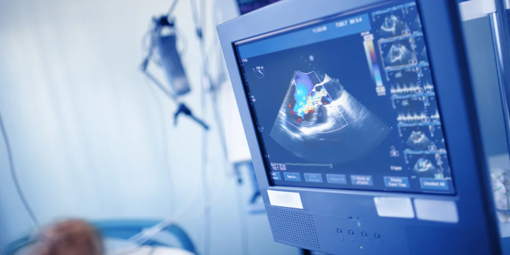
This method is frequently employed to “observe” the actual heart structural action. The computer displays a cone-shaped 2-D echo perspective that shows the heart’s structural movements in real time. Our doctor can examine and assess the various cardiac structures thanks to this technology.
Our doctors utilize 2D Echo to comprehensively assess cardiac function, evaluate the effectiveness of both surgical and medical treatments, and detect conditions such as heart valve diseases, myocardial disorders, pericardial diseases, congenital heart anomalies, and cardiac tumors.”
Our 2D Echo services include plain imaging, color Doppler with Doppler, carotid Doppler, peripheral studies, and renal index measurements.
What Our Patients Say
Hear from our valued patients about their experiences at Chirayu Super Speciality Hospital and how our care has made a positive impact on their health and well-being.


Grateful for Chirayu Hospital’s 2D Echo team. They provided a detailed assessment of my heart health with care and professionalism.

Chirayu Hospital’s 2D Echo service exceeded my expectations. They offered thorough insights and compassionate care.

The 2D Echo procedure at Chirayu Hospital was seamless. They provided comprehensive insights into my heart health.

The 2D Echo service at Chirayu Hospital was efficient and detailed. They answered all my questions and eased my concerns.

Impressed by the professionalism of Chirayu Hospital’s 2D Echo technicians. They made my heart examination comfortable and informative.

Chirayu Hospital’s 2D Echo team ensured accurate results and compassionate care during my heart assessment.
Meet Our Medical Specialists
Our experts at Chirayu Super Speciality Hospital provide precise cardiac evaluations using advanced technology for effective treatment and heart health.


Frequently Asked Questions
Here, we provide answers to some of the most commonly asked questions to help you better understand our services, policies, and facilities. If you have any additional questions, please do not hesitate to contact us.
A 2D Echo (Two-Dimensional Echocardiogram) is a non-invasive test that uses sound waves to create images of the heart, allowing specialists to assess its structure and function.
A 2D Echo is performed to diagnose and monitor heart conditions, evaluate heart function, and detect abnormalities in the heart’s structure.
Individuals experiencing symptoms like chest pain, shortness of breath, or irregular heartbeats, or those with a history of heart disease, should consider a 2D Echo.
A 2D Echo uses ultrasound waves to produce images of the heart. A transducer is placed on the chest, emitting sound waves that bounce off the heart structures and create detailed images.
Yes, a 2D Echo is a safe and painless procedure with no known risks or side effects.